

How to prevent diseases for dairy cows
Disorders of the Claw Capsule in Cattle
Etiology and Pathogenesis
Sinking of the claw due to activation of matrix metalloproteinase, which is a feature of subclinical laminitis, is the major predisposing factor. As the space between the flexor process of the pedal bone narrows, the corium is crushed, causing ischemic necrosis and compromising horn production. This results in a hole forming in the sole. As the damaged corium undergoes repair, granulation tissues erupt through the hole in the sole. Because this condition is strongly associated with subclinical laminitis, some softening of the sole horn will occur. This increases the rate of horn wear. Softening of the sole horn also occurs under very unhygienic conditions when horn is exposed to the fluid component in slurry.
Iatrogenic forms of the lesion are produced when inexperienced claw trimmers remove too much horn from beneath the heel, resulting in abnormal pressure on the dermis. Excessive wear of the softened sole horn flattens and thins the sole.
Heel erosion is another potential contributing cause of a sole ulcer. Normally, weight is borne by the bulb of the heel, but if heel erosion occurs, weight-bearing may be transferred forward to the region beneath the flexor process. Sometimes, a displaced pad of horn slips over to the vulnerable area, causing abnormal pressure over the flexor process of the distal phalanx.
Clinical Findings
The progress and severity of lameness are variable and often masked in bilateral cases, depending on the size of the lesion and extent of the secondary infection. As the lateral hind digit is usually involved, the limb is often held slightly abducted with weight-bearing on the unaffected medial digit. In tie stalls, the hind toes may be rested on the edge of a curb in an attempt to relieve pressure on the heel-sole junction. On flat surfaces, an affected animal stands with the hindlimbs camped back. Some cows may shake the affected foot frequently, while those with bilateral lesions may continually shift weight from limb to limb and frequently lie down.
The earliest stages of this lesion may be discovered during routine claw trimming. As sole horn is removed from over the typical site, a hemorrhagic lesion may be exposed. The clinical lesion varies from a soft, slightly discolored area that may be painful under pressure to a distinct bright red knob of granulation tissue. This is often the stage at which lameness becomes severe enough to be noticed. Once the corium is exposed, infection can invade the deeper structures of the claw and spread proximally to involve the navicular bursa, resulting in necrosis of the flexor tendon and ligaments of the navicular bone. A retroarticular abscess may develop, which may be further complicated by infection of the distal interphalangeal joint. Rupture of the flexor tendon leads to dorsal rotation (upward) of the toe (“cocked toe”). In complicated cases, infection may progress up the deep flexor tendon sheath.


Treatment
Treatment must be aimed at removing pressure from the affected area. Skillful therapeutic claw trimming is highly effective. This procedure lowers the entire bearing surface of the lateral claw, which transfers loading to the sound medial claw. Applying a “lift” has become the accepted treatment for this condition. The simplest form of lift is a wooden or rubber block that is glued or nailed to the unaffected medial claw, thereby removing all weight-bearing from the ulcer region. Recently, various models of easier-to-apply plastic slippers have been developed. Care must be taken when applying either a block or a slipper to avoid the sharp hard rear edge of the device from causing pressure under the sole. Blocks should be removed at ∼1 mo to avoid causing damage to the sole.
Protruding granulation tissue need not be excised and must not be treated with any caustic agent, as this can retard healing. Bandages should not be applied because this results in continued weight-bearing at the ulcer site. Furthermore, covering the lesion causes it to remain moist and promotes maceration and bacterial infection.
Many ulcers never fully resolve, and affected cows may have chronic low-grade lameness and need corrective foot trimming 2–4 times/yr for their productive lives.
Prevention and Control
Because the development of sole ulcers is intimately related to subclinical laminitis (p 974), the latter should be investigated and appropriate control measures instituted.
White Line Disease
As with sole ulcers, white line disease commonly affects one or both lateral hind claws, predominantly in heavy, high-yielding dairy cattle kept under confined conditions. The condition is characterized by hemorrhage into or separation (avulsion) of the white line on the apical or abaxial border of the sole, but most commonly at the heel-sole junction. The corium becomes infected through this lesion.
Etiology and Pathogenesis
The white line is an extension of the lamellae and is composed of soft horn joining the sole to the wall. White line disease is believed to be strongly associated with subclinical laminitis. It is postulated that stretching of collagen fibers, combined with sinking of the pedal bone, accounts for the hemorrhage into the white line that is so frequently observed.
Rupture of the white line is exacerbated by the impact of locomotion, particularly among animals housed on concrete. The abaxial region of the wall of the hindlimb is the area of the claw absorbing the concussion of the first impact of locomotion.
Solid foreign bodies may lodge in the softened, widened zone. They may push through to the corium beneath and introduce infection; however, the presence of a foreign body is not essential for the lesion to develop.
There are 3 possible sequelae of localized infection: 1) a localized abscess may develop; 2) infection may be forced proximally along the spaces between the lamellae to form a track that may discharge at the coronary band; 3) the infected track may, as it forces its way proximally, infect other structures, depending on the site of the initial infection. An anterior track can infect the distal interphalangeal joint directly. Tracks forming closer to the heel are likely to cause infection of the bursa of the deep flexor tendon. Invariably, the bursa ruptures into the retroarticular space, and an abscess develops in this location. Infection of the distal interphalangeal joint and the tendon sheath of the deep flexor tendon may follow. Necrosis and avulsion of the insertion of the deep flexor tendon into the distal phalanx are frequent complications.
Clinical Findings
The lateral claw of the hindfoot (often both) is usually involved. If bilateral, the disease may remain unnoticed until lameness is more pronounced in one limb than the other. Because the outer hind claw is affected, the limb is swung away from the body during each stride. The animal may stand with the medial claw bearing weight. White line separation without complications is frequently seen at claw trimming. The degree of pain and lameness depends on the rate of development and extent of the subsolar abscess. Routine examination of the sole must include the complete exploration of the abaxial white line region. Black marks must be explored with the tip of a hoof knife as potential sites for track formation. Discharge of pus from the skin/horn junction above the abaxial wall is always reason to suspect a white line lesion. In these cases, the white line must always be examined very carefully.


Swelling of the heel bulb represents the most advanced form of this condition; it is frequently misdiagnosed as footrot (often presented as a case of footrot that is resistant to treatment). Footrot causes the whole foot to swell evenly to the fetlock; in contrast, a retroarticular abscess leads to enlargement of only one heel bulb.
Treatment
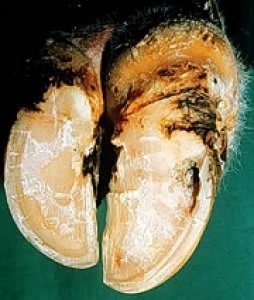
During a claw examination, any black mark in the white line must be cut out until healthy horn is exposed. For a local abscess, removal of an elliptical segment of the wall adjacent to the lesion aids free drainage by providing a self-cleansing abaxial opening. Cream-colored pus may indicate a corporeal response to tissues tearing as collagen fibers stretch and the pedal bone sinks. In contrast, if the pus is black, it is likely that infection has penetrated from the outside.
Abscessation with sinus formation at the coronary band requires the removal of a segment of the abaxial wall (∼0.75 cm wide) from the white line to the coronary band. This procedure is best performed with the cutting disk of a grinding tool under local anesthesia. Often, a plug of necrotic debris is found in the track.
Retroarticular abscesses are usually quite large and surrounded by a mass of fibroelastic tissue that inhibits drainage. Drainage is accomplished by passing a probe through the abscess from the lesion on the abaxial wall until it can be palpated under the skin on the axial surface of the bulb. An incision is made onto the probe and a drainage tube is drawn through the abscess. Continuous irrigation of the lesion for several days with saline is indicated. The application of a lift to the sound claw is helpful, as is complete immobilization of the digit. Immobilization of the joint reduces the risk of permanent deformity due to avulsion of the deep flexor tendon.
Toe Ulcer
Toe ulcer is the term used to describe any hemorrhagic lesion of the dermis occurring in the apical region of the sole and/or white line, most frequently in the lateral hind claw.
Etiology and Pathogenesis
Recent discussion has revealed 3 distinct etiopathologies that can produce a similar clinical appearance: 1) As subclinical laminitis progresses, in some cases the distal phalanx will rotate. Stretching of the collagen fibers and movement of the bone will cause tearing of tissues, including the circumferential artery. Hemorrhage will result. In extreme instances, the tip of the bone will prolapse through the apex of the sole. Many animals with a rotated digit also have a ridge (the reaction ridge) running around the wall. The ridge is similar in location to a hardship groove and is displaced distally in a similar manner. Osteomyelitis of the distal phalanx can be seen in complicated cases. 2) Many cases have been reported (anecdotally) in which the anterior half of the sole has been worn down almost paper thin. Hemorrhage from bruising is seen through the thin horn at the apex. Breakdown of the horn and formation of an abscess have been reported. The probable cause is a painful lesion in the heel, which forces the animal to throw most of its weight onto the anterior part of the sole. 3) Necrosis of the apex of the pedal bone is extremely common in yearling beef calves after transportation over long distances. The condition is sporadically reported in mature cows. In either case, it is suspected that standing for long periods is the cause. The main blood supply to the digit is the very large axial digital artery which could be vulnerable to pressure. This artery connects with the terminal arch that penetrates across the bone. Necropsy reveals that necrosis occurs distal to this artery, and radiographs tend to suggest the same thing. The terminal artery possibly marks the margin at which pathologic fractures of the distal digit can be observed.
Clinical Findings
In many cases, the white line and sole in the toe region may be stained with serum or blood. In more advanced cases, a prolapse of the sole may occur with associated infection. The exact nature of the lesion probably cannot be determined without radiography.


Treatment and Control
In mature cattle in which the lesion has resulted from wear and no complications are obvious, the cavity should be cleansed, dried, packed with an antibiotic powder, and covered with methyl methacrylate. If the bottom of the lesion is black in these cattle, a probe should be inserted; if necrotic tissue can be detected, l–2 cm of the apex of the toe should be removed with hoof cutters. The condition of the pedal bone should be visible. If necrosis of the bone can be confirmed, regional anesthesia should be applied and a further l–2 cm of toe removed. If the wound bleeds profusely, it is likely that necrosis is not extensive. When hemorrhage is minimal, it is probable that necrosis of the bone is extensive or a physiologic fracture is present. There are several reports of the toe (not the claw) having been amputated with satisfactory recovery. Systemic antibiotics and application of a lift to the sound claw is advised. If the wound is obviously contaminated, the lesion should be packed with a hygroscopic mixture (50% magnesium sulfate and 50% glycerin) and bandaged for a maximum of 24 hr, after which the lesion should be thoroughly dried, dressed with antibiotic powder, and closed with methyl methacrylate. If rotation of the digit has been detected radiographically, the prognosis for recovery is poor.
Control of subclinical laminitis is likely to lower the incidence of toe ulcer.
The incidence of apical necrosis can be quite high in young feedlot cattle after they have been transported over long distances. Many of these cattle become recumbent and die from pneumonia. The lesion can be treated in the same manner as for mature cattle, but it is probably not cost effective to do so.
Double Sole
In this condition, a superficial sole is separated by a space from a second sole beneath.

Etiology and Pathogenesis
The cause is unknown. It is postulated that a sudden disturbance in the microcirculation of the dermis probably results in an effusion of serum separating the dermis from the epidermis. Double sole has been seen in cattle suddenly changed from a mainly forage diet to one rich in concentrates and in beef cattle turned out in the spring on lush grass after a winter ration of forage. It has also been observed in a herd that was forced to walk on hard surfaces after claw trimming. Double sole can be confused with underunning of the heel which is a frequent sequela of white line disease.
Treatment and Control
Treatment is simple unless mismanaged. The abaxial wall must remain completely intact and only a portion of the sole covering the bulb cut away. The sole beneath is extremely soft and vulnerable to damage; therefore, the animal should be confined to a well-strawed loose stall until the new horn has hardened, after which more of the sole may be removed.
Sudden changes in the quality of the forage should be avoided. Double sole has also been observed after feeding moldy hay.
Foreign Bodies in the Sole
Occasionally, a foreign body such as a stone, chip of glass, or nail becomes embedded in the sole. Even if the material does not penetrate to the corium, localized pressure causes pain and lameness. Removal of the foreign body usually resolves the lameness without incident.


If the foreign body penetrates through to the corium, infection is introduced to the dermal level and an abscess develops. The rapidity of onset and severity of the lameness depends to some extent on the location of the sole penetration.
In the apical and subapical region, the abscess is located between the distal phalanx and the nonresilient sole. As the abscess develops, interungular pressure increases rapidly. Thus, the onset of lameness is rapid and the degree of pain very severe. Acute lameness may cause the animal to stand with the foot off the ground or with the toe lightly touching. A differential diagnosis is fracture of the distal phalanx.
Treatment consists of removing the foreign body and coring out the track to the corium with a fine-pointed hoof knife. Creating a large hole is inappropriate. Pus is often released under considerable pressure. Antibiotic should be squeezed into the cavity, which closes rapidly. The opening should not be plugged but covered with elastic waterproof material to prevent blockage with mud or manure.
In the sub-bulbar region, the corium is located between the digital cushion (a flexible structure) and the soft resilient horn of the bulb.
The onset of lameness is relatively slow, and the pain generated is significant but not severe. The pus in the abscess tends to spread over a wide area through the fascial plane and to cause separation of the skin-horn junction at the heel. A moist discharge from this area may be the first indication of the lesion. This is referred to as underrunning of the heel, a condition that can be confused with double sole (see above).
Treatment consists of removing the foreign body if still present. The detached horn should not be stripped off in its entirety. Part of the detached horn may be removed, but the abaxial wall must be left intact to bear weight and spare the exposed newly forming sole. Bandaging may not be required, but the animal should be housed in a well-strawed area for a few days.
Sandcracks
(Vertical fissures)
Sandcracks are vertical fissures or cracks in the wall of the claw. They account for ∼0.2% of lesions of the claws of dairy cows. In western Canada, the average incidence in mature beef cows is ∼20%. In individual herds, the incidence can be as high as 60%. No breed differences have been recorded. The lesion is extremely unsightly, which can be a considerable drawback for beef cattle producers who wish to sell their animals.


Clinical Findings
Vertical fissures occur almost exclusively in the lateral fore claw. They can start at the coronary band and run part or all of the way to the bearing surface. In herds with the highest incidence of this disorder, lesions are observed to start from a horizontal groove in the middle of the claw.
Etiology
The etiology remains uncertain. The incidence is highest in mature, heavy cows. Bending or buckling of the claw around one or several horizontal grooves probably creates mechanical stresses causing the rupture of the claw wall.
Treatment
Most sandcracks are not painful and require no treatment. However, if the origin of the lameness can be traced to a claw in which a sandcrack is present, routine treatment of the crack is appropriate.
A crack at the coronary band can split open and infection can enter. The protrusion of granulation tissue through such a lesion can be quite troublesome. The dorsal pouch of the distal interphalangeal joint is extremely superficial at this point. The joint is very vulnerable, and lesions at this location should never be ignored. Superficial horn should be pared away and an astringent dressing applied (50% mixture of a sulfa powder and anhydrous copper sulfate). Pressure should be applied using cotton batten held tightly in place by a narrow adhesive elastic bandage encircling the entire coronary band.
The fissures often have ragged edges that may be twisted and gape open. Sometimes the edges move and pick up a foreign body, which can cause trouble. Cosmetic treatment may be requested in the case of show animals. The axial wall at the tip of the claw should be cut back so that the weight is borne only by the abaxial portion of the wall. The ragged edges of the fissure should be trimmed, ideally with the cutting disk of a grinding tool. In selected cases, a fissure can be immobilized with an application of methyl methacrylate after the 2 edges of the fissure have been laced together with steel wire.
Horizontal Fissures
Horizontal fissures result from disruption of horn production at the dermis beneath the coronary band, leading to a defect in the integrity of the wall. These fissures run parallel to the coronary band. The defect varies in severity from a shallow groove (hardship groove) to a complete fracture (fissure) of the wall. A comparable anomaly is seen as a band of horn differing in appearance from the remainder of the claw. One form of the band is seen in animals stressed following weaning (weaning groove) or during a period of nutritional deprivation. The fissure moves distally as the claw grows, and the distal portion becomes progressively more mobile (thimble) until it fractures, leaving a “broken toe.” A series of grooves can destabilize the vertical strength of the dorsal wall causing it to bend (buckled toe).
Etiology
Fissures are believed to be caused by a wide variety of stressors, including a bad calving, an acute febrile disease, or a sudden, relatively short-term but significant change in nutrition. The pathophysiology of a groove or fissure is likely to be a short-term laminitis-like event, but it does not necessarily indicate the onset of subclinical laminitis (see Subclinical Laminitis). A depressed band may indicate a period of stress such as would follow weaning. A ridge may indicate an event such as compensatory growth.
Clinical Findings
The horizontal groove or fissure is an important indicator of metabolic disturbance. The date on which the causal insult occurred can be calculated by measuring the distance from the hair line to the fissure and dividing that number by the growth rate of the claw. In mature dairy cows, the rate of growth of the wall measured along the dorsal flexure of the claw is ∼0.5 cm/mo. Growth rates are more rapid in young animals, in animals on intensive feed, and during the summer months.


Treatment
Most cases require no treatment. Very deep fissures may eventually result in the formation of a thimble, which is extremely painful. In these cases, the loose horn should be removed with pincers; regional anesthesia may be needed.
Corkscrew Claw
A corkscrew claw is twisted throughout its length in a configuration that displaces the abaxial wall by up to 360°. One or both lateral hind claws may be affected in cows >4 yr old. Although corkscrew claws are rarely seen in bulls, many believe there is a heritable component.

Pathogenesis
Bone molding is seen in the distal phalanx, but it is not known if this is a matter of cause or effect. Periarticular exostoses develop around the distal interphalangeal joint, possibly resulting from strain of the distal abaxial collateral ligament. Pressure from the exostosis on the dermis of the wall probably accounts for the excessive growth of the abaxial wall.
Treatment
Correctly trimming a corkscrew claw requires much skill. The horn formation is extremely hard and difficult to cut. The abnormally narrow shape of the distal phalanx makes it difficult to pare the claw without causing bleeding at the toe. The strategy is to shorten the claw as much as possible without causing bleeding. Next, the horn wall that is displaced beneath the claw is cut away. Then, so far as is possible, the horn is shaped to approximate normal. Trimming helps the animal get around for a while, but does not “cure” the condition. Affected animals should ultimately be culled.
Slipper Foot
A slipper foot is named for its alleged likeness to a Persian slipper. The claw is flat and curled upward to form a square end. The horn is heavily ridged and has lost its shine, and the coronary band is rougher and darker than normal. Although there is no objective evidence to support the theory, the slipper foot is probably synonymous with chronic laminitis and may be a sequela of either acute or subclinical laminitis. Treatment is always disappointing. The claw can be shaped to approximate normal, but invariably it collapses and serious sequelae follow. Animals with slipper foot should be culled as soon as economically appropriate.

Last full review/revision March 2012 by Paul R. Greenough, FRCVS
![]()